A stable scan isn't one thing.
The abdominal imaging literature, read for you. RadBrief EP10.
▶ Prefer to listen? Hit play above. Same read, about 4 minutes.
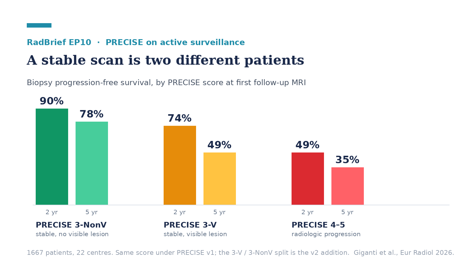
PRECISE has been the recommended way to report serial MRI in prostate active surveillance since 2017, and until now it rested almost entirely on expert consensus. This is the first international multicenter outcome data behind it, 1667 patients across 22 centers, and the system holds up: radiologic progression carried 4.53-fold higher odds of biopsy progression than a stable or regressing scan. But the finding I’d actually use is more specific: a stable scan splits into two very different patients depending on whether there was a visible lesion to begin with.
It’s a retrospective study of men on surveillance for GG 1 or GG 2 disease, scans from December 2005 to July 2022, with biopsy histopathology as the reference standard. Local expert radiologists re-reported 4597 scans. One detail worth knowing before you read the abstract: those readers scored on PRECISE v1, because the re-reads happened in 2021 and 2022 and v2 wasn’t published until 2024. The v2 subdivision of PRECISE 3 was reconstructed afterward from baseline lesion visibility, which works because that information was already in the record. So this validates the v1 scale and supports one piece of v2. The rest of v2, the PI-QUAL quality gate, PRECISE X, the standardized two-diameter measurement, isn’t tested here, and the authors don’t claim otherwise.
What I’d tell a resident. Learn the split. Under v1 a stable scan was a single category, PRECISE 3, and v2 divides it into 3-V, stable with a visible lesion, and 3-NonV, stable with nothing visible. The five-year progression-free rates were 49 percent for 3-V and 78 percent for 3-NonV. Same score, same word “stable” in the report, and roughly a thirty-point spread in outcome. The 3-NonV patients behaved about like the ones whose lesions had actually regressed, while at five years the 3-V group was doing about as poorly as the radiologic-progression group had been doing at two. I’d also score against both the baseline scan and the most recent prior, not just the last one, because a lesion can be stable since the previous study and clearly larger than where it started.
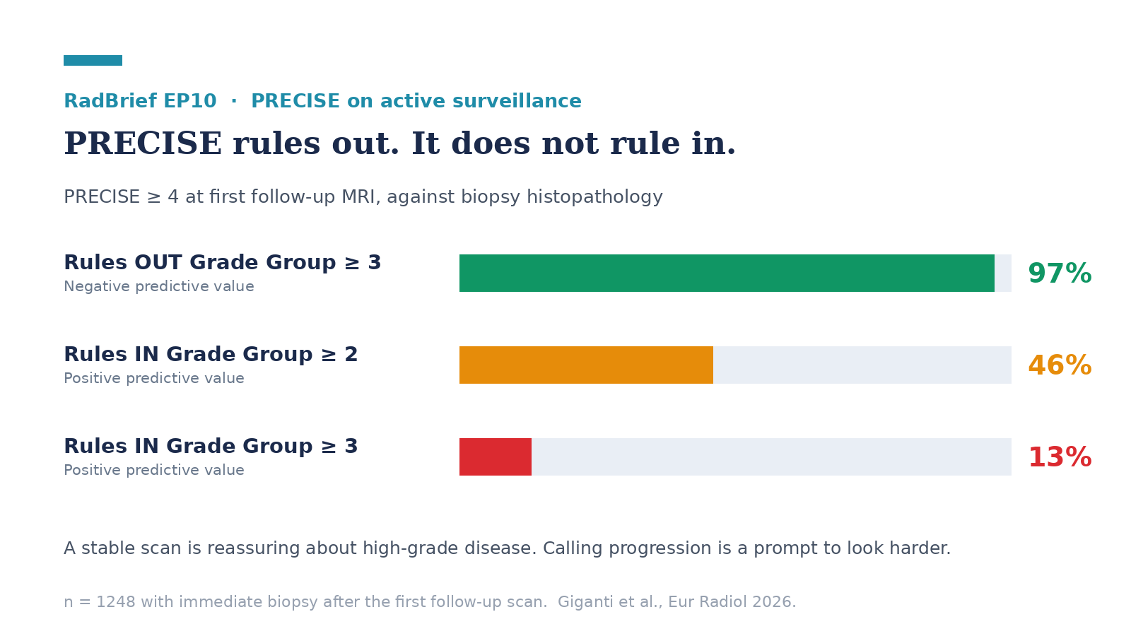
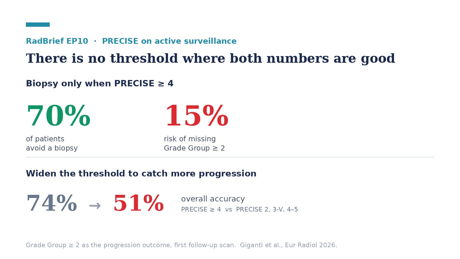
What I’d tell a general radiologist. The value of PRECISE is in the negative, and I think it’s worth being precise about which negative. For ruling out GG ≥ 3 disease the negative predictive value was 97 percent, so a stable scan is genuinely reassuring about high-grade cancer, and that’s what supports dropping protocol biopsies. Calling progression is a much weaker signal: positive predictive value was 46 percent for GG ≥ 2 and 13 percent for GG ≥ 3. So I’d report PRECISE 4 or 5 as a reason to look harder and to talk to urology, not as a statement that the cancer has progressed. The trade-off is quantified, which I appreciate. Biopsy only for PRECISE ≥ 4 and about 70 percent of patients avoid a biopsy, at roughly a 15 percent risk of missing GG ≥ 2. Widen the threshold to catch more and overall accuracy drops from 74 percent to 51 percent. There’s no setting where both numbers are good, and the paper is honest about that.
What we should be skeptical about. There was no central image review and no predefined protocol for what counts as radiologic progression. The authors cite their own 2026 multicenter paper showing high inter- and intra-rater variability in lesion measurement during surveillance, which cuts directly against the reproducibility this system depends on, and I think that’s the real open question rather than whether the scale predicts anything. Second, the biopsy decisions weren’t blind to the MRI. Urologists chose who got biopsied, often influenced by the same scan being evaluated, so the progression rates carry ascertainment bias. Third, poor-quality scans were excluded by local subjective judgment rather than a formal quality score, and the authors concede that some of what was scored as progression may be image quality improving rather than the tumor changing. That’s a real problem for a system whose v2 now formally gates on PI-QUAL, and it’s untested here. Two smaller things: the negative predictive value decays across serial scans, 85 percent at the first follow-up down to 79 and then 71, so the reassurance is strongest early. And the biparametric subgroup was 47 patients against 1201, which is too small to tell us whether PRECISE works without contrast.
Worth noting the Cleveland Clinic was one of the participating centers, with Ryan Ward as a co-author and Andrei Purysko among the surveillance initiative collaborators.
The thing this study can’t address is the one v2 leaned on hardest, which is scan quality. PI-QUAL is the next piece, and I’ll come back to it.
I’m Melina Pectasides, and this is RadBrief.
Reference block: what changed from PRECISE v1 to v2
Screenshot this. It’s the five things that actually differ.
Scan quality
v1: not addressed
v2: PI-QUAL ≥ 4 required for a valid score
Baseline scan
v1: no formal definition
v2: the first MRI linked to the cancer diagnosis, pre- or post-biopsy
Score 3
v1: one category
v2: split into 3-V (visible lesion, stable) and 3-NonV (non-visible, stable)
New category
v1: none
v2: PRECISE X, when quality is too poor to score
Measurement
v1: not standardized
v2: two diameters on T2, ideally matching the prior scan
Three more v2 specifics I’d keep in mind
Minimum one year between scans to call a significant change
A volume increase over 50 percent is the suggested threshold for significant growth
A new PI-RADS or Likert ≥ 3 lesion forces the scan to 4 or 5, even if everything else is stable
The numbers behind the episode
1667 patients, 22 centers, 4597 scans; 90 percent GG 1 at entry; median follow-up 4 years
First follow-up scan: 11 percent regression, 62 percent stable, 27 percent progression
PRECISE 4–5 vs 1–3, adjusted OR for biopsy progression 4.53 (95% CI 3.37–6.12)
PRECISE ≥ 4 for GG ≥ 2: sensitivity 57, specificity 79, PPV 46, NPV 85, accuracy 74
PRECISE ≥ 4 for GG ≥ 3: NPV 97, PPV 13
NPV across serial scans: 85 → 79 → 71 percent
Deeper reading
The paper: Giganti F, Leni R, Wagaskar V, et al. Multicentre validation of the PRECISE scoring system for prostate MRI during active surveillance. European Radiology. 2026. https://doi.org/10.1007/s00330-026-12570-z
The original system: Moore CM, Giganti F, Albertsen P, et al. Reporting Magnetic Resonance Imaging in Men on Active Surveillance for Prostate Cancer: The PRECISE Recommendations. European Urology. 2017;71:648–655. https://doi.org/10.1016/j.eururo.2016.06.011
The update: Englman C, Maffei D, Allen C, et al. PRECISE Version 2: Updated Recommendations for Reporting Prostate MRI in Patients on Active Surveillance. European Urology. 2024. https://doi.org/10.1016/j.eururo.2024.03.014
How to actually apply v2: Ogata A, Brembilla G, Dias AB, et al. PRECISE Version 2: Essential Tips for Prostate Cancer Monitoring Using MRI. RadioGraphics. 2026;46(5):e250126. https://doi.org/10.1148/rg.250126
Why the reproducibility worry is real: Englman C, Adebusoye B, Cosenza M, et al. Inter- and intra-rater variability of MRI-based lesion size measurements in active surveillance for prostate cancer: a multicentre study. European Radiology. 2026. https://doi.org/10.1007/s00330-025-12318-1
The prior evidence base: Rajwa P, Pradere B, Quhal F, et al. Reliability of Serial Prostate MRI to Detect Prostate Cancer Progression During Active Surveillance: A Systematic Review and Meta-analysis. European Urology. 2021;80:549–563. https://doi.org/10.1016/j.eururo.2021.05.001